Login
Subscribe
Infographic: Drivers of the Expansion of Volume Electron Microscopy
Infographic: Drivers of the Expansion of Volume Electron Microscopy
Technological advancements over the last two decades transformed volume electron microscopy, improving usability, resolution, and throughput.
Technological advancements over the last two decades transformed volume electron microscopy, improving usability, resolution, and throughput.
electron microscopy

The Expansion of Volume Electron Microscopy
Danielle Gerhard, PhD | Sep 8, 2023 | 6 min read
A series of technological advancements for automation and parallel imaging made volume electron microscopy more user friendly while increasing throughput.

Cryo-EM: Building on a History of Invention and Innovation
Thermo Fisher Scientific | Aug 2, 2023 | 1 min read
From humble yet ingenious beginnings to Nobel recognition, cryogenic electron microscopy (cryo-EM) provides insights into scientific questions that other technologies are unable to answer.

Form Determines Function: Insights from Structural Biology
The Scientist’s Creative Services Team | 1 min read
Researchers use diverse tools to analyze protein structures.

How Fear Restructures the Mouse Brain
Natalia Mesa, PhD | Aug 15, 2022 | 4 min read
By combining deep learning and electron microscopy, researchers now have a more detailed understanding of how fear changes the brain.

What’s the Deal with Bacterial Nanotubes?
Sruthi S. Balakrishnan | Jun 1, 2021 | 10+ min read
Several labs have reported the formation of bacterial nanotubes under different, often contrasting conditions. What are these structures and why are they so hard to reproduce?

Transforming Virology Research with Cryo-EM
The Scientist’s Creative Services Team and Thermo Fisher Scientific | 1 min read
Explore what researchers can do with Cryo-EM

Infographic: Sources of Variation in Bacterial Nanotube Studies
Sruthi S. Balakrishnan | Jun 1, 2021 | 2 min read
Differences in how researchers prepare and image samples can lead to discrepancies in their results.

Coronavirus Closeup, 1964
Ashley Yeager | Sep 1, 2020 | 3 min read
Electron microscopy revealed that a deadly disease of birds was not a form of flu, but a different type of virus entirely.
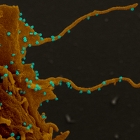
Coronavirus-Infected Cells Grow Filopodia
Shawna Williams | Jun 30, 2020 | 1 min read
SARS-CoV-2 causes cells to put out projections that spread the virus, a study finds.

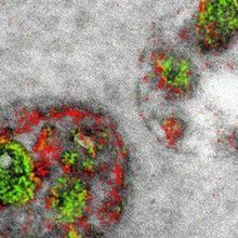
Image of the Day: Coronavirus Under the Scope
Amy Schleunes | Feb 17, 2020 | 1 min read
The National Institute of Allergy and Infectious Diseases releases a series of images that offer a close up look at the novel coronavirus SARS-CoV-2.

Imaging Chromatin to Deduce Function from Form
Marissa Fessenden | Dec 1, 2018 | 7 min read
Researchers describe their tools for probing how the physical shape of the genome affects genes’ function.
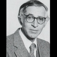
Aaron Klug, Developer of Crystallographic Electron Microscopy, Dies
Ashley P. Taylor | Nov 26, 2018 | 3 min read
The chemist and biophysicist won a Nobel prize for the development of a technique to probe the structures of nucleotide-protein complexes.

Image of the Day: Feather Mites
The Scientist Staff and The Scientist Staff | Feb 19, 2018 | 1 min read
Researchers used scanning electron microscopy to peer at bugs on several hummingbird species.

Image of the Day: Beetle Penis
The Scientist Staff and The Scientist Staff | Dec 22, 2017 | 1 min read
Scientists look to a leaf beetle’s genitals for lessons on improving catheter strength.

Scientists Who Developed Cryo-Electron Microscopy Win Nobel Prize
Diana Kwon | Oct 4, 2017 | 3 min read
Chemistry Nobel goes to Jacques Dubochet, Joachim Frank, and Richard Henderson.

Entire Fruit Fly Brain Imaged with Electron Microscopy
Ashley Yeager | May 31, 2017 | 3 min read
Synaptic connections and a new neuron type emerge in high-res images, which hold promise for mapping the complete connectome.

Doors and Pores
Mary Beth Aberlin | Dec 1, 2016 | 2 min read
The awesome architecture of the gateways to the nucleus
Electron Micrographs Get a Dash of Color
Ben Andrew Henry | Nov 3, 2016 | 4 min read
A new technique creates colorful stains that label proteins and cellular structures at higher resolution than ever before possible.

Microscopy’s Growth Through the Years
Jenny Rood | Oct 1, 2016 | 5 min read
From confocal fluorescence microscopy to super-resolution and live 3-D imaging, microscopes have changed rapidly since 1986.